DICOTYLEDONOUS ROOT
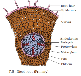
In the primary section of root, there are 3 distinct regions namely epidermis, cortex and stele.
Epidermis (epiblema or piliferous layer)
$\displaystyle \small \circ$ It is the outer most single layer of tissue.
$\displaystyle \small \circ$ It consist compactly arranged thin walled parenchyma without intercellular space.
$\displaystyle \small \circ$ The cells are barrel shaped, some epidermal cells have unicellular extensions called root hairs or trichoblast.
$\displaystyle \small \circ$ The cuticle is absent over epidermis.
$\displaystyle \small \circ$ The root hairs are living and perform all vital functions, these are involved in water absorption.
$\displaystyle \small \circ$ The epidermis is also called epiblema or rhizodermis.
Cortex
$\displaystyle \small \circ$ It lies below the epiblema and occupies major portion of the root anatomy.
$\displaystyle \small \circ$ It has two zones the outer general cortex and inner endodermis.
$\displaystyle \small \circ$ The general cortex is the massive region consist several layers of thin walled parenchyma with intercellular space.
$\displaystyle \small \circ$ These are homogeneous cells which stores starch.
$\displaystyle \small \circ$ The endodermis is the innermost layer cortex, which is made up of thick walled parenchyma.
$\displaystyle \small \circ$ These cells cover the stellar region.
$\displaystyle \small \circ$ The endodermis is thick walled by the casparin strips, it is formed by deposition of lignin.
$\displaystyle \small \circ$ The lignin is deposited in transverse wall only, it is absent in tangential wall.
$\displaystyle \small \circ$ No inner tangential wall thickenings in endodermis, hence all endoderm cells are passage cells.
Stele
$\displaystyle \small \circ$ It is also called vascular cylinders, it consist pericycle, vascular bundles and pith.
$\displaystyle \small \circ$ The pericycle occur below the endodermis. It is made up of single layer of parenchyma cells. These can undergo cell division and produces lateral branches.
$\displaystyle \small \circ$ The vascular bundles are radial type, the xylem and phloem are in separate groups. There are four patch of xylem and they alternates with phloem, it is called tetrarch vascular bundles. In the xylem the smaller protoxylem are towards margin, which develop centripetal called exarch.
$\displaystyle \small \circ$ The space between vascular bundle is filled with thin walled parenchyma called conjunctive tissue. The pith is generally absent in dicot root, if present it is negligible.

MONOCOTYLEDONOUS ROOT
In the anatomy of Monocot root, the T.S. of maize root shows there are 3 distinct regions namely epidermis, cortex and stele.
Epidermis
$\displaystyle \small \circ$ It is the outer most region made up of single layer of compactly arranged parenchyma cells.
$\displaystyle \small \circ$ It is also called epiblema or piliferous layer.
$\displaystyle \small \circ$ The cuticle and stomata are absent.
$\displaystyle \small \circ$ The number of unicellular extensions are present on epidermis, called root hairs, which helps in absorption of water.
Cortex
$\displaystyle \small \circ$ It is the second region lies inside the epidermis.
$\displaystyle \small \circ$ It consist several layers of thin walled parenchyma with intercellular space.
$\displaystyle \small \circ$ In older roots few layers of parenchyma beneath epidermis become thick walled by deposition of subarin. It forms exodermis.
$\displaystyle \small \circ$ The general cortex consist thin walled parenchyma.
$\displaystyle \small \circ$ The endodermis is the inner most layer of cortex.
$\displaystyle \small \circ$ The endodermal cells are transversely covered by lignin form casparin strips.
$\displaystyle \small \circ$ In addition to its presence, lignin and cellulose are arranged in ‘U’ shaped thickenings.
$\displaystyle \small \circ$ The cells of endodermis are impermeable, hence some cells just opposite to the protoxylem are thin walled which forms passage cells. Such cells allow water to enter.
$\displaystyle \small \circ$ The passage cells are also called transfusion cells.
Stele
$\displaystyle \small \circ$ It is the inner region composed of pericycle, vascular bundles and pith.
$\displaystyle \small \circ$ The pericycle is single layer of thin walled parenchyma, it does not have any function.
$\displaystyle \small \circ$ The vascular bundle consist several patches of xylem and phloem hence it is polyarch. The vascular bundles are radial type.
$\displaystyle \small \circ$ The space between xylem and phloem is filled with thin walled parenchyma called conjunctive tissue. The xylem is exarch in nature, which develop centripetally. The pith is prominent in monocot root. It stores starch and reserve food.

DICOTYLEDONOUS STEM
The internal structure of stem can be studied by taking the T.S.of young dicot stem that shows 3 distinct regions namely epidermis, cortex and stele.
Epidermis
$\displaystyle \small \circ$ It is the outer most region.
$\displaystyle \small \circ$ It consist single layer of barrel shaped living cells, and they are compactly arranged without intercellular space.
$\displaystyle \small \circ$ The epidermal cells develop multicellular hairs called trichome.
$\displaystyle \small \circ$ The epidermis is covered by non-living layer cuticle, the presence of openings in epidermis called stomata, which are covered by guard cells.
Cortex
$\displaystyle \small \circ$ It is next to the epidermis and is differentiated into hypodermis, general cortex and endodermis.
$\displaystyle \small \circ$ The hypodermis is beneath the epidermis, it consist few layers of collenchyma.
$\displaystyle \small \circ$ The cell wall is thick at corners by the deposition of pectin hence intercellular space is absent.
$\displaystyle \small \circ$ These are living cells with protoplasm.
$\displaystyle \small \circ$ The hypoderm provides mechanical support.
$\displaystyle \small \circ$ The general cortex is next to the hypodermis, it consist several layers of thin walled parenchyma with intercellular space.
$\displaystyle \small \circ$ These cells contain chloroplast hence they are chlorenchyma.
$\displaystyle \small \circ$ This region consist few cells secretes latex gum etc., these are called resin canals.
$\displaystyle \small \circ$ The endodermis is the innermost region of cortex.
$\displaystyle \small \circ$ It consist barrel shaped cells, they are compactly arranged without inter cellular space.
$\displaystyle \small \circ$ It is wavy in nature, the cells contain starch hence it is also called as starch sheath.
Stele
$\displaystyle \small \circ$ The tissue lies inside endodermis is the stele.
$\displaystyle \small \circ$ It consist pericycle, medullary rays, vascular bundle and pith.
$\displaystyle \small \circ$ The pericycle is the outer region of stele, it consist parenchyma and the sclerenchyma in alternate arrangement.
$\displaystyle \small \circ$ The sclerenchyma is found outside vascular bundle which is like cap hence it is called as bundle cap.
$\displaystyle \small \circ$ The medullary rays are thin walled parenchyma found between vascular bundles, it is connected with pith.
$\displaystyle \small \circ$ The pith is the central part which is in storage function.
$\displaystyle \small \circ$ The vascular bundles are numerous and arranged in discontinuous rings hence it is called eustele.
$\displaystyle \small \circ$ Each bundle is conjoint, collateral and open type.
$\displaystyle \small \circ$ Phloem is towards margin and xylem is towards centre, in between xylem and phloem there is presence of cambium called intrafascicular cambium.
$\displaystyle \small \circ$ The protoxylem is towards inner side hence it is endarch.

MONOCOTYLEDONOUS STEM
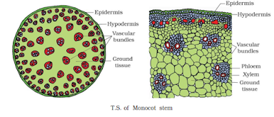
The T.S. of Maize stem at inter nodal region shows two distinct regions namely epidermis and ground tissue containing vascular bundles.
Epidermis
$\displaystyle \small \circ$ It is the outer zone, which consist a single layer of compactly arranged barrel shaped cells.
$\displaystyle \small \circ$ The epidermis is covered by non-living cuticle.
$\displaystyle \small \circ$ The stem hairs are absent because the inter node is covered by sheathing leaf base.
Ground tissue
$\displaystyle \small \circ$ In monocot stem the tissue inside epidermis is ground tissue but it can’t be differentiated.
$\displaystyle \small \circ$ Just below the epidermis, presence of 2-3 layers of thick walled dead sclerenchyma forms hypodermis, which provides mechanical support.
$\displaystyle \small \circ$ The rest of the section consist thin walled parenchyma with inter cellular space, it is called ground tissue.
$\displaystyle \small \circ$ The vascular bundles are scattered in the ground tissue.
Vascular bundle
$\displaystyle \small \star$ These are not arranged in the ring, but they are scattered in the ground tissue.
$\displaystyle \small \star$ The vascular bundles are numerous, they are smaller towards margin and larger towards centre.
$\displaystyle \small \star$ Each bundle is oval shaped, which is covered by sclerenchyma forms bundle sheath.
$\displaystyle \small \star$ The vascular bundles are conjoint, collateral and closed type.
$\displaystyle \small \star$ The xylem appears in “Y” shape, there are two larger metaxylem and one or two protoxylem.
$\displaystyle \small \star$ The excess protoxylem join to form a larger cavity called lysigenous cavity or water cavity.
$\displaystyle \small \star$ The phloem is towards margin, the xylem is exarch in nature and space between xylem and phloem is occupied by sclerenchyma.

DORSIVENTRAL (DICOTYLEDONOUS) LEAF
$\displaystyle \small \bullet$ The leaves are with broad flat surface carry out function of photosynthesis and transpiration.
$\displaystyle \small \bullet$ It has two surfaces, the upper surface is called adaxial surface and lower surface is called aboxial surface.
$\displaystyle \small \bullet$ In dicot leaves mesophyll is differentiated as upper palisade parenchyma and the lower spongy parenchyma, hence these are also called dorsiventral leaf.
$\displaystyle \small \bullet$ The C.S. of sunflower leaf shows 3 distinct arrangements namely epidermis, mesophyll and vascular bundle.
Epidermis
$\displaystyle \small \circ$ The epidermis cover both upper and lower surface called as upper epidermis and lower epidermis.
$\displaystyle \small \circ$ Both are made up of single layer of flattened parenchyma cells, they are compactly arranged and without chloroplast.
$\displaystyle \small \circ$ The epidermis is covered by thick waxy, non-living layer called cuticle and presence of multicellular leaf hairs trichome.
$\displaystyle \small \circ$ In the lower epidermis presence of small openings called stomatal pores, each pore is surrounded by specialized guard cells with chloroplast.
$\displaystyle \small \circ$ The pore and guard cells forms stomata, they help in gaseous exchange.
$\displaystyle \small \circ$ In dorsiventral or dicot leaf stomata are present on lower surface hence the leaf is described as hypostomic leaf.
Mesophyll
$\displaystyle \small \circ$ The tissue located between two epidermis in leaf is the mesophyll, it is differentiated into two regions.
$\displaystyle \small \circ$ The palisade parenchyma is towards adaxile surface, it consist 2 layers of elongated, cylindrical cells of parenchyma. They are arranged right angle with narrow intercellular space. The cells are with abundant chloroplast.
$\displaystyle \small \circ$ The spongy parenchyma is towards aboxile surface. It consist loosely arranged cells with large inter cellular space. The cells are irregular shaped and sponge like. The cells are with chloroplast, the main functions are transpiration, gaseous exchange and photosynthesis.
Vascular bundle
$\displaystyle \small \circ$ These are located in midrib and in veins.
$\displaystyle \small \circ$ Each bundle is conjoint, collateral and closed, the bundles are covered by thin walled parenchyma forms bundle sheath.
$\displaystyle \small \circ$ In midrib region the space between epidermis and vascular bundle is filled with thick walled collenchyma which provides support.
$\displaystyle \small \circ$ The collenchyma and parenchyma forms bundle sheath extensions.
$\displaystyle \small \circ$ In the vascular bundle, xylem is towards adaxile side, it consist tracheids, vessels, fibres and xylem parenchyma.
$\displaystyle \small \circ$ The phloem is towards aboxile surface it has sieve tubes, companion cells and parenchyma.

ISOBILATERAL (MONOCOTYLEDONOUS) LEAF
In the monocot leaf the adaxile and aboxile surface are similar in their external and internal structure, hence it is also called as Isobilateral leaf.
The cross section of maize leaf shows there are 3 distinct regions namely epidermis, mesophyll and vascular bundle.
Epidermis
$\displaystyle \small \circ$ It is found on both the surface, the upper epidermis and lower epidermis, they are similar in structure.
$\displaystyle \small \circ$ Each epidermis is made up of single layer of compactly arranged barrel shaped cells.
$\displaystyle \small \circ$ They are externally covered by cuticle and presence of multicellular hairs trichome.
$\displaystyle \small \circ$ The stomata are found in both upper and lower epidermis, hence it is called amphistomic leaf.
$\displaystyle \small \circ$ In upper epidermis there are few large , thin walled cells called motor cells or bulliform cells they helps wilting or rolling.
Mesophyll
$\displaystyle \small \circ$ The mesophyll is between upper and lower epidermis.
$\displaystyle \small \circ$ It is homogeneous and consist the thin walled, isodiametric, living parenchyma cells with inter cellular space.
$\displaystyle \small \circ$ They have chloroplast, the mesophyll is not differentiated and it is photosynthesis in function.
Vascular bundle
$\displaystyle \small \circ$ The vascular bundles are at veins.
$\displaystyle \small \circ$ The parallel veins represent vascular bundles.
$\displaystyle \small \circ$ These are covered by parenchyma with starch granules.
$\displaystyle \small \circ$ The bundles are connected to upper and lower epidermis by sclerenchyma, it provides mechanical support.
$\displaystyle \small \circ$ The vascular bundles are conjoint, collateral and closed type.
$\displaystyle \small \circ$ The xylem is endarch; usually there are two metaxylem and few protoxylem.
$\displaystyle \small \circ$ Xylem is towards upper epidermis and the phloem is towards lower epidermis.
$\displaystyle \small \circ$ The vascular bundles are surrounded by enlarged parenchyma with organized chloroplast.
$\displaystyle \small \circ$ This type of bundle sheath cell arrangement is called Kranz anatomy.

In the primary section of root, there are 3 distinct regions namely epidermis, cortex and stele.
Epidermis (epiblema or piliferous layer)
$\displaystyle \small \circ$ It is the outer most single layer of tissue.
$\displaystyle \small \circ$ It consist compactly arranged thin walled parenchyma without intercellular space.
$\displaystyle \small \circ$ The cells are barrel shaped, some epidermal cells have unicellular extensions called root hairs or trichoblast.
$\displaystyle \small \circ$ The cuticle is absent over epidermis.
$\displaystyle \small \circ$ The root hairs are living and perform all vital functions, these are involved in water absorption.
$\displaystyle \small \circ$ The epidermis is also called epiblema or rhizodermis.
Cortex
$\displaystyle \small \circ$ It lies below the epiblema and occupies major portion of the root anatomy.
$\displaystyle \small \circ$ It has two zones the outer general cortex and inner endodermis.
$\displaystyle \small \circ$ The general cortex is the massive region consist several layers of thin walled parenchyma with intercellular space.
$\displaystyle \small \circ$ These are homogeneous cells which stores starch.
$\displaystyle \small \circ$ The endodermis is the innermost layer cortex, which is made up of thick walled parenchyma.
$\displaystyle \small \circ$ These cells cover the stellar region.
$\displaystyle \small \circ$ The endodermis is thick walled by the casparin strips, it is formed by deposition of lignin.
$\displaystyle \small \circ$ The lignin is deposited in transverse wall only, it is absent in tangential wall.
$\displaystyle \small \circ$ No inner tangential wall thickenings in endodermis, hence all endoderm cells are passage cells.
Stele
$\displaystyle \small \circ$ It is also called vascular cylinders, it consist pericycle, vascular bundles and pith.
$\displaystyle \small \circ$ The pericycle occur below the endodermis. It is made up of single layer of parenchyma cells. These can undergo cell division and produces lateral branches.
$\displaystyle \small \circ$ The vascular bundles are radial type, the xylem and phloem are in separate groups. There are four patch of xylem and they alternates with phloem, it is called tetrarch vascular bundles. In the xylem the smaller protoxylem are towards margin, which develop centripetal called exarch.
$\displaystyle \small \circ$ The space between vascular bundle is filled with thin walled parenchyma called conjunctive tissue. The pith is generally absent in dicot root, if present it is negligible.

MONOCOTYLEDONOUS ROOT
In the anatomy of Monocot root, the T.S. of maize root shows there are 3 distinct regions namely epidermis, cortex and stele.
Epidermis
$\displaystyle \small \circ$ It is the outer most region made up of single layer of compactly arranged parenchyma cells.
$\displaystyle \small \circ$ It is also called epiblema or piliferous layer.
$\displaystyle \small \circ$ The cuticle and stomata are absent.
$\displaystyle \small \circ$ The number of unicellular extensions are present on epidermis, called root hairs, which helps in absorption of water.
Cortex
$\displaystyle \small \circ$ It is the second region lies inside the epidermis.
$\displaystyle \small \circ$ It consist several layers of thin walled parenchyma with intercellular space.
$\displaystyle \small \circ$ In older roots few layers of parenchyma beneath epidermis become thick walled by deposition of subarin. It forms exodermis.
$\displaystyle \small \circ$ The general cortex consist thin walled parenchyma.
$\displaystyle \small \circ$ The endodermis is the inner most layer of cortex.
$\displaystyle \small \circ$ The endodermal cells are transversely covered by lignin form casparin strips.
$\displaystyle \small \circ$ In addition to its presence, lignin and cellulose are arranged in ‘U’ shaped thickenings.
$\displaystyle \small \circ$ The cells of endodermis are impermeable, hence some cells just opposite to the protoxylem are thin walled which forms passage cells. Such cells allow water to enter.
$\displaystyle \small \circ$ The passage cells are also called transfusion cells.
Stele
$\displaystyle \small \circ$ It is the inner region composed of pericycle, vascular bundles and pith.
$\displaystyle \small \circ$ The pericycle is single layer of thin walled parenchyma, it does not have any function.
$\displaystyle \small \circ$ The vascular bundle consist several patches of xylem and phloem hence it is polyarch. The vascular bundles are radial type.
$\displaystyle \small \circ$ The space between xylem and phloem is filled with thin walled parenchyma called conjunctive tissue. The xylem is exarch in nature, which develop centripetally. The pith is prominent in monocot root. It stores starch and reserve food.

DICOTYLEDONOUS STEM
The internal structure of stem can be studied by taking the T.S.of young dicot stem that shows 3 distinct regions namely epidermis, cortex and stele.
Epidermis
$\displaystyle \small \circ$ It is the outer most region.
$\displaystyle \small \circ$ It consist single layer of barrel shaped living cells, and they are compactly arranged without intercellular space.
$\displaystyle \small \circ$ The epidermal cells develop multicellular hairs called trichome.
$\displaystyle \small \circ$ The epidermis is covered by non-living layer cuticle, the presence of openings in epidermis called stomata, which are covered by guard cells.
Cortex
$\displaystyle \small \circ$ It is next to the epidermis and is differentiated into hypodermis, general cortex and endodermis.
$\displaystyle \small \circ$ The hypodermis is beneath the epidermis, it consist few layers of collenchyma.
$\displaystyle \small \circ$ The cell wall is thick at corners by the deposition of pectin hence intercellular space is absent.
$\displaystyle \small \circ$ These are living cells with protoplasm.
$\displaystyle \small \circ$ The hypoderm provides mechanical support.
$\displaystyle \small \circ$ The general cortex is next to the hypodermis, it consist several layers of thin walled parenchyma with intercellular space.
$\displaystyle \small \circ$ These cells contain chloroplast hence they are chlorenchyma.
$\displaystyle \small \circ$ This region consist few cells secretes latex gum etc., these are called resin canals.
$\displaystyle \small \circ$ The endodermis is the innermost region of cortex.
$\displaystyle \small \circ$ It consist barrel shaped cells, they are compactly arranged without inter cellular space.
$\displaystyle \small \circ$ It is wavy in nature, the cells contain starch hence it is also called as starch sheath.
Stele
$\displaystyle \small \circ$ The tissue lies inside endodermis is the stele.
$\displaystyle \small \circ$ It consist pericycle, medullary rays, vascular bundle and pith.
$\displaystyle \small \circ$ The pericycle is the outer region of stele, it consist parenchyma and the sclerenchyma in alternate arrangement.
$\displaystyle \small \circ$ The sclerenchyma is found outside vascular bundle which is like cap hence it is called as bundle cap.
$\displaystyle \small \circ$ The medullary rays are thin walled parenchyma found between vascular bundles, it is connected with pith.
$\displaystyle \small \circ$ The pith is the central part which is in storage function.
$\displaystyle \small \circ$ The vascular bundles are numerous and arranged in discontinuous rings hence it is called eustele.
$\displaystyle \small \circ$ Each bundle is conjoint, collateral and open type.
$\displaystyle \small \circ$ Phloem is towards margin and xylem is towards centre, in between xylem and phloem there is presence of cambium called intrafascicular cambium.
$\displaystyle \small \circ$ The protoxylem is towards inner side hence it is endarch.

MONOCOTYLEDONOUS STEM
The T.S. of Maize stem at inter nodal region shows two distinct regions namely epidermis and ground tissue containing vascular bundles.
Epidermis
$\displaystyle \small \circ$ It is the outer zone, which consist a single layer of compactly arranged barrel shaped cells.
$\displaystyle \small \circ$ The epidermis is covered by non-living cuticle.
$\displaystyle \small \circ$ The stem hairs are absent because the inter node is covered by sheathing leaf base.
Ground tissue
$\displaystyle \small \circ$ In monocot stem the tissue inside epidermis is ground tissue but it can’t be differentiated.
$\displaystyle \small \circ$ Just below the epidermis, presence of 2-3 layers of thick walled dead sclerenchyma forms hypodermis, which provides mechanical support.
$\displaystyle \small \circ$ The rest of the section consist thin walled parenchyma with inter cellular space, it is called ground tissue.
$\displaystyle \small \circ$ The vascular bundles are scattered in the ground tissue.
Vascular bundle
$\displaystyle \small \star$ These are not arranged in the ring, but they are scattered in the ground tissue.
$\displaystyle \small \star$ The vascular bundles are numerous, they are smaller towards margin and larger towards centre.
$\displaystyle \small \star$ Each bundle is oval shaped, which is covered by sclerenchyma forms bundle sheath.
$\displaystyle \small \star$ The vascular bundles are conjoint, collateral and closed type.
$\displaystyle \small \star$ The xylem appears in “Y” shape, there are two larger metaxylem and one or two protoxylem.
$\displaystyle \small \star$ The excess protoxylem join to form a larger cavity called lysigenous cavity or water cavity.
$\displaystyle \small \star$ The phloem is towards margin, the xylem is exarch in nature and space between xylem and phloem is occupied by sclerenchyma.

DORSIVENTRAL (DICOTYLEDONOUS) LEAF
$\displaystyle \small \bullet$ The leaves are with broad flat surface carry out function of photosynthesis and transpiration.
$\displaystyle \small \bullet$ It has two surfaces, the upper surface is called adaxial surface and lower surface is called aboxial surface.
$\displaystyle \small \bullet$ In dicot leaves mesophyll is differentiated as upper palisade parenchyma and the lower spongy parenchyma, hence these are also called dorsiventral leaf.
$\displaystyle \small \bullet$ The C.S. of sunflower leaf shows 3 distinct arrangements namely epidermis, mesophyll and vascular bundle.
Epidermis
$\displaystyle \small \circ$ The epidermis cover both upper and lower surface called as upper epidermis and lower epidermis.
$\displaystyle \small \circ$ Both are made up of single layer of flattened parenchyma cells, they are compactly arranged and without chloroplast.
$\displaystyle \small \circ$ The epidermis is covered by thick waxy, non-living layer called cuticle and presence of multicellular leaf hairs trichome.
$\displaystyle \small \circ$ In the lower epidermis presence of small openings called stomatal pores, each pore is surrounded by specialized guard cells with chloroplast.
$\displaystyle \small \circ$ The pore and guard cells forms stomata, they help in gaseous exchange.
$\displaystyle \small \circ$ In dorsiventral or dicot leaf stomata are present on lower surface hence the leaf is described as hypostomic leaf.
Mesophyll
$\displaystyle \small \circ$ The tissue located between two epidermis in leaf is the mesophyll, it is differentiated into two regions.
$\displaystyle \small \circ$ The palisade parenchyma is towards adaxile surface, it consist 2 layers of elongated, cylindrical cells of parenchyma. They are arranged right angle with narrow intercellular space. The cells are with abundant chloroplast.
$\displaystyle \small \circ$ The spongy parenchyma is towards aboxile surface. It consist loosely arranged cells with large inter cellular space. The cells are irregular shaped and sponge like. The cells are with chloroplast, the main functions are transpiration, gaseous exchange and photosynthesis.
Vascular bundle
$\displaystyle \small \circ$ These are located in midrib and in veins.
$\displaystyle \small \circ$ Each bundle is conjoint, collateral and closed, the bundles are covered by thin walled parenchyma forms bundle sheath.
$\displaystyle \small \circ$ In midrib region the space between epidermis and vascular bundle is filled with thick walled collenchyma which provides support.
$\displaystyle \small \circ$ The collenchyma and parenchyma forms bundle sheath extensions.
$\displaystyle \small \circ$ In the vascular bundle, xylem is towards adaxile side, it consist tracheids, vessels, fibres and xylem parenchyma.
$\displaystyle \small \circ$ The phloem is towards aboxile surface it has sieve tubes, companion cells and parenchyma.

ISOBILATERAL (MONOCOTYLEDONOUS) LEAF
In the monocot leaf the adaxile and aboxile surface are similar in their external and internal structure, hence it is also called as Isobilateral leaf.
The cross section of maize leaf shows there are 3 distinct regions namely epidermis, mesophyll and vascular bundle.
Epidermis
$\displaystyle \small \circ$ It is found on both the surface, the upper epidermis and lower epidermis, they are similar in structure.
$\displaystyle \small \circ$ Each epidermis is made up of single layer of compactly arranged barrel shaped cells.
$\displaystyle \small \circ$ They are externally covered by cuticle and presence of multicellular hairs trichome.
$\displaystyle \small \circ$ The stomata are found in both upper and lower epidermis, hence it is called amphistomic leaf.
$\displaystyle \small \circ$ In upper epidermis there are few large , thin walled cells called motor cells or bulliform cells they helps wilting or rolling.
Mesophyll
$\displaystyle \small \circ$ The mesophyll is between upper and lower epidermis.
$\displaystyle \small \circ$ It is homogeneous and consist the thin walled, isodiametric, living parenchyma cells with inter cellular space.
$\displaystyle \small \circ$ They have chloroplast, the mesophyll is not differentiated and it is photosynthesis in function.
Vascular bundle
$\displaystyle \small \circ$ The vascular bundles are at veins.
$\displaystyle \small \circ$ The parallel veins represent vascular bundles.
$\displaystyle \small \circ$ These are covered by parenchyma with starch granules.
$\displaystyle \small \circ$ The bundles are connected to upper and lower epidermis by sclerenchyma, it provides mechanical support.
$\displaystyle \small \circ$ The vascular bundles are conjoint, collateral and closed type.
$\displaystyle \small \circ$ The xylem is endarch; usually there are two metaxylem and few protoxylem.
$\displaystyle \small \circ$ Xylem is towards upper epidermis and the phloem is towards lower epidermis.
$\displaystyle \small \circ$ The vascular bundles are surrounded by enlarged parenchyma with organized chloroplast.
$\displaystyle \small \circ$ This type of bundle sheath cell arrangement is called Kranz anatomy.


0 Comments