CONNECTIVE TISSUES
This tissue connects different tissues and different organs.
Connective tissues are most abundant and widely distributed in the body of complex animals.
Characteristics:
1. The cells are not in groups, they are freely distributed in inter cellular spaces.
2. The intercellular spaces are filled with non-living material called matrix or ground substance. The matrix is secreted by cells.
3. The cells and fibres are suspended in the matrix.
4. Theses tissues have own blood vassals called vascularized tissues.
5. These are derived from mesoderm.
6. These tissues undergo regeneration by producing new cells which helps in healing of wounds.
Classification
i) Loose connective tissue
ii) Dense connective tissue
iii) Specialized connective tissue
Loose connective tissue
Cells and fibres are loosely arranged in a semi-fluid ground substance.
Types of Loose connective tissues
i) Areolar tissue
ii) Adipose tissue
iii) Reticular tissue
Areolar connective tissues
$\displaystyle \small \bullet$ Most widely distributed connective tissue in the body.
$\displaystyle \small \bullet$ Areolar tissue connects integuments with muscles.
$\displaystyle \small \bullet$ It is called loose connective tissue, which is located below epithelium, around the organs.
$\displaystyle \small \bullet$ These tissues fill the space in the organs.
$\displaystyle \small \bullet$ This tissue is composed of cells, fibres and matrix.
 $\displaystyle \small \bullet$ Types of fibres in the matrix
i) White fibres are also called collagen fibres, they are found in bundles made up of protein collagen. They are hard and rigid. These are unbranched and runs in all directions.
ii) Yellow fibres are also called elastic fibres. They occur single made up of protein elastin. They are branched and show elasticity.
iii) Reticular fibres are generally invisible. They are thin branching fibres, forms network and resemble the white fibres.
$\displaystyle \small \bullet$ Types of cells in the matrix
i) Fibroblast are larger, irregular branching cells. They produce and maintain the fibres.
ii) Adipocytes or fat cells are round flat cells. They store a drop of lipid.
iii) Macrophages are large moving cells called amoebocytes. They destroy foreign bodies and serve as defensive function.
iv) Mast cells are large cells with spherical nucleus, which secretes matrix, heparin and histamine.
v) Plasma cells are smaller oval cells with small nucleus. They produce antibodies.
Adipose tissues
$\displaystyle \small \bullet$ The loose connective tissue which consists large number of fat cells or adipocytes are called adipose tissues.
$\displaystyle \small \bullet$ Each cells have large vacuole, containing a drop of lipid(fat). Hence the cytoplasm and nucleus are pushed towards the margin.
$\displaystyle \small \bullet$ This tissue contains mast cells, reticulate fibre and very little matrix.
$\displaystyle \small \bullet$ This tissue is found below the skin, around kidney, buttocks etc.
$\displaystyle \small \bullet$ The increase of adipose tissue deposited around the trunk forms obesity.
$\displaystyle \small \bullet$ It helps in storage and release of fat.
$\displaystyle \small \bullet$ It serves as the shock absorber under the skin.
$\displaystyle \small \bullet$ It helps to conserve the body heat.

Reticular Tissues
$\displaystyle \small \bullet$ The cells are in star shape and form network like structure.
$\displaystyle \small \bullet$ These are present in spleen, lymph nodes etc.
$\displaystyle \small \bullet$ They are helpful in formation of lymphoid tissues in the body.
$\displaystyle \small \bullet$ In the bone marrow, they help in formation of blood cells.
Dense connective tissue
$\displaystyle \small \bullet$ They contain cells, fibres and fibroblasts which are compactly packed.
$\displaystyle \small \bullet$ The orientation of fibres may be regular or irregular pattern.
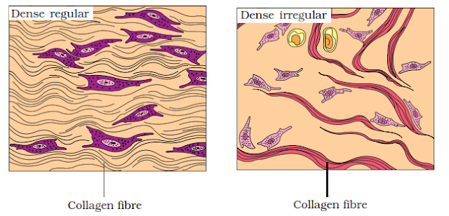
$\displaystyle \small \bullet$ In dense regular connective tissues, collagen fibres are present in rows between parallel bundles of fibres as in tendons and ligaments.
$\displaystyle \small \bullet$ Tendons attach skeletal muscles to bones. They are made up of white fibrous tissues. Tendons are tough and inelastic.
$\displaystyle \small \bullet$ Ligaments attach one bone to another bone. They are made up of yellow elastic tissues. they are strong and elastic.
$\displaystyle \small \bullet$ Dense irregular connective tissue has fibroblasts and many fibres (mostly collagen), that are arranged differently. These tissues are present in the skin.

Specialized connective tissue
Connective tissues that are specialized for specific function are called specialized connective tissues.
Types of specialized connective tissues
i) Cartilage
ii) Bones
iii) Blood
Cartilage
$\displaystyle \small \bullet$ This tissue is strong and elastic but smoother than bone.
$\displaystyle \small \bullet$ The elasticity is due to the presence of protein chondrin.
$\displaystyle \small \bullet$ The matrix is solid, pliable and resists compression.
$\displaystyle \small \bullet$ Cells of this tissue (chondrocytes) are enclosed in small cavities within the matrix secreted by them.
$\displaystyle \small \bullet$ Most of the cartilages in vertebrate embryos are replaced by bones in adults.
$\displaystyle \small \bullet$ Types of Cartilage
i) Hyaline cartilage: It is semi rigid, semitransparent with thin bluish tint. It is made up of matrix and cells. The hyaline cartilage is found at the tip of bones, treachea, larynx etc.
ii) Elastic cartilage: It is structurally almost similar to hyaline cartilage. It is covered by the membrane perichondrium and it is made up of matrix, cells and fibres. This is found at the tip of nose, external ear pinna and epiglottis.
iii) Fibro cartilage: This cartilage does not consist perichondrium. It consist large number of white fibres in matrix. This is found at regions of great compression which gives cushion effect. It is found in inter vertebral disc.

Bone
$\displaystyle \small \bullet$ Bones have a hard and non-pliable ground substance rich in calcium salts and collagen fibres.
$\displaystyle \small \bullet$ It is the main tissue that provides structural frame to the body.
$\displaystyle \small \bullet$ Bones support and protect softer tissues and organs.
$\displaystyle \small \bullet$ The matrix consists of longitudinal canals called Haversian canals.
$\displaystyle \small \bullet$ Limb bones, such as the long bones of the legs, serve weight-bearing functions.
$\displaystyle \small \bullet$ They also interact with skeletal muscles attached to them to bring about movements.
$\displaystyle \small \bullet$ The bone marrow in some bones is the site of production of blood cells.

Blood
$\displaystyle \small \bullet$ Blood is a fluid connective tissue.
$\displaystyle \small \bullet$ It transports gases, nutrient and metabolic wastes.
$\displaystyle \small \bullet$ It carries hormones, maintain the Osmotic pressure, PH of blood and regulates body temperature.
$\displaystyle \small \bullet$ Components of blood:
i) Plasma: Plasma is the fluid part of blood without form elements. It is light straw coloured containing water, organic and inorganic substances.
ii) Corpuscles
a. RBC (Red Blood Cells): They are small, enucleated, spherical, biconcave. Haemoglobin is present inside RBC.
b. WBC (White Blood Cells): Larger than RBC, less in number, colourless and nucleated.
Types of WBC
1. Agranulocytes: Cytoplasm is non granular.
$\displaystyle \small \star$ Lymphocytes (20-25%): Formed in lymph node and are smallest WBC associated with antibody production. Nucleus is round and large.
$\displaystyle \small \star$ Monocytes (3-8%): Large cells with kidney shaped or horseshoe shaped nucleus. They are motile and phagocytic in function.
2. Granulocyte: Cytoplasm is granular.
$\displaystyle \small \star$ Eosinophils (2-4%): WBC stains deep red with acidic dyes. The nucleus is bilobed.
$\displaystyle \small \star$ Basophils (0.5-1.5%): Stains blue with basic stains. Nucleus is lobed and irregular shaped cytoplasmic granule.
$\displaystyle \small \star$ Neutrophils(60-70%): Cells stain violet with neutral dyes. Nucleus is multilobed with connected thin chromotic strands.
iii) Platelets (Thrombocytes): These are only fragments. They are non-nucleated, round and small in size. Life span is about 5-9 days. They initiate blood clotting.
MUSCLE TISSUES
$\displaystyle \small \bullet$ The muscle consist long, cylindrical fibres called fibrils, which are also called as myofibrils.
$\displaystyle \small \bullet$ The cells of this tissue are known for their function of contraction and relaxation.
$\displaystyle \small \bullet$ The cells are long hence called as muscle fibres.
$\displaystyle \small \bullet$ The cell has the cytoplasm called sarcoplasm and their cell membrane is called sarcolemma.
$\displaystyle \small \bullet$ Types of muscular tissues
i) Smooth muscles or unstriated muscle
ii) Striated or skeletal muscles
iii) Cardiac muscles
Smooth muscles
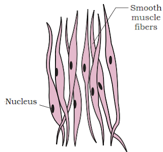
$\displaystyle \small \bullet$ This tissue cells are spindle shaped and tapers at both the ends.
$\displaystyle \small \bullet$ Each cell is covered by thin sarcolemma and it consist sarcoplasm.
$\displaystyle \small \bullet$ The cross striations are absent hence it also called as non-striated muscle.
$\displaystyle \small \bullet$ Single distinct nucleus is present in the middle of the cell.
$\displaystyle \small \bullet$ Cell junctions hold them and are bundled together in a connective tissue sheath.
$\displaystyle \small \bullet$ The wall of internal organs such as the blood vessels, stomach and intestine contains this type of muscle tissue.
$\displaystyle \small \bullet$ Smooth muscles are ‘involuntary’, as their functioning cannot be directly controlled.
 Skeletal muscles
$\displaystyle \small \bullet$ They are also known as striated, voluntary muscles.
$\displaystyle \small \bullet$ They are closely attached to the skeletal bones, hence are called skeletal muscles.
$\displaystyle \small \bullet$ Each cell consists outer covering sarcolemma, which is thick and tough, forming a sheath.
$\displaystyle \small \bullet$ It consist the cytoplasm called sarcoplasm and many nuclei.
$\displaystyle \small \bullet$ The nuclei are peripheral and directly attach to sarcolemma.
$\displaystyle \small \bullet$ In sacroplasm the myofibrils are composed of actin and myosin, which appears in alternate dark and light bands.
$\displaystyle \small \bullet$ It is voluntary in function and it undergoes quick contraction when suffered by fatigue.

Cardiac muscles
$\displaystyle \small \bullet$ Cardiac muscle tissue is a contractile tissue, present only in the heart.
$\displaystyle \small \bullet$ Cell junctions fuse the plasma membranes of cardiac muscle cells and make them stick together.
$\displaystyle \small \bullet$ Communication junctions (intercalated discs) at some fusion points allow the cells to contract as a unit.
$\displaystyle \small \bullet$ The cells are cylindrical and branched, which forms network.
$\displaystyle \small \bullet$ They have sarcolemma, sarcoplasm and are uninucleated.
$\displaystyle \small \bullet$ Cardiac muscles are involuntary, as their functioning cannot be directly controlled.

NEURAL TISSUES
$\displaystyle \small \bullet$ The nervous system has control over the body’s responsiveness to changing conditions.
$\displaystyle \small \bullet$ This tissue is responsible for conduction of impulses, and is derived from ectoderm.
$\displaystyle \small \bullet$ There are two types of cells in nervous tissue, they are neuron and neuroglea cells.
$\displaystyle \small \bullet$ Neuron is a structural and functional unit of nervous system consisting cyton, dendrites and axon.
$\displaystyle \small \bullet$ When a neuron is suitably stimulated, an electrical disturbance is generated which swiftly travels along its plasma membrane.
$\displaystyle \small \bullet$ Arrival of the disturbance at the neuron’s endings or output zone triggers events that may cause stimulation or inhibition of adjacent neurons and other cells.
$\displaystyle \small \bullet$ There are four types of neuroglea cells namely: astrocytes, microglia, oligodendrites and epidermal cells.

This tissue connects different tissues and different organs.
Connective tissues are most abundant and widely distributed in the body of complex animals.
Characteristics:
1. The cells are not in groups, they are freely distributed in inter cellular spaces.
2. The intercellular spaces are filled with non-living material called matrix or ground substance. The matrix is secreted by cells.
3. The cells and fibres are suspended in the matrix.
4. Theses tissues have own blood vassals called vascularized tissues.
5. These are derived from mesoderm.
6. These tissues undergo regeneration by producing new cells which helps in healing of wounds.
Classification
i) Loose connective tissue
ii) Dense connective tissue
iii) Specialized connective tissue
Loose connective tissue
Cells and fibres are loosely arranged in a semi-fluid ground substance.
Types of Loose connective tissues
i) Areolar tissue
ii) Adipose tissue
iii) Reticular tissue
Areolar connective tissues
$\displaystyle \small \bullet$ Most widely distributed connective tissue in the body.
$\displaystyle \small \bullet$ Areolar tissue connects integuments with muscles.
$\displaystyle \small \bullet$ It is called loose connective tissue, which is located below epithelium, around the organs.
$\displaystyle \small \bullet$ These tissues fill the space in the organs.
$\displaystyle \small \bullet$ This tissue is composed of cells, fibres and matrix.

i) White fibres are also called collagen fibres, they are found in bundles made up of protein collagen. They are hard and rigid. These are unbranched and runs in all directions.
ii) Yellow fibres are also called elastic fibres. They occur single made up of protein elastin. They are branched and show elasticity.
iii) Reticular fibres are generally invisible. They are thin branching fibres, forms network and resemble the white fibres.
$\displaystyle \small \bullet$ Types of cells in the matrix
i) Fibroblast are larger, irregular branching cells. They produce and maintain the fibres.
ii) Adipocytes or fat cells are round flat cells. They store a drop of lipid.
iii) Macrophages are large moving cells called amoebocytes. They destroy foreign bodies and serve as defensive function.
iv) Mast cells are large cells with spherical nucleus, which secretes matrix, heparin and histamine.
v) Plasma cells are smaller oval cells with small nucleus. They produce antibodies.
| White fibres(collagen) | Yellow fibres(Elastic) |
|---|---|
| These are white in colour | These are yellow in colour |
| These are thick and more in number | These are thin and few in number. |
| Wavy and unbranched | Straight and branched. |
| Hard and rigid | Elastic in nature |
| Made up of protein collagen | Made up of protein elastin |
| Always occur in bundle | Occur in single |
Adipose tissues
$\displaystyle \small \bullet$ The loose connective tissue which consists large number of fat cells or adipocytes are called adipose tissues.
$\displaystyle \small \bullet$ Each cells have large vacuole, containing a drop of lipid(fat). Hence the cytoplasm and nucleus are pushed towards the margin.
$\displaystyle \small \bullet$ This tissue contains mast cells, reticulate fibre and very little matrix.
$\displaystyle \small \bullet$ This tissue is found below the skin, around kidney, buttocks etc.
$\displaystyle \small \bullet$ The increase of adipose tissue deposited around the trunk forms obesity.
$\displaystyle \small \bullet$ It helps in storage and release of fat.
$\displaystyle \small \bullet$ It serves as the shock absorber under the skin.
$\displaystyle \small \bullet$ It helps to conserve the body heat.

Reticular Tissues
$\displaystyle \small \bullet$ The cells are in star shape and form network like structure.
$\displaystyle \small \bullet$ These are present in spleen, lymph nodes etc.
$\displaystyle \small \bullet$ They are helpful in formation of lymphoid tissues in the body.
$\displaystyle \small \bullet$ In the bone marrow, they help in formation of blood cells.
Dense connective tissue
$\displaystyle \small \bullet$ They contain cells, fibres and fibroblasts which are compactly packed.
$\displaystyle \small \bullet$ The orientation of fibres may be regular or irregular pattern.
$\displaystyle \small \bullet$ In dense regular connective tissues, collagen fibres are present in rows between parallel bundles of fibres as in tendons and ligaments.
$\displaystyle \small \bullet$ Tendons attach skeletal muscles to bones. They are made up of white fibrous tissues. Tendons are tough and inelastic.
$\displaystyle \small \bullet$ Ligaments attach one bone to another bone. They are made up of yellow elastic tissues. they are strong and elastic.
$\displaystyle \small \bullet$ Dense irregular connective tissue has fibroblasts and many fibres (mostly collagen), that are arranged differently. These tissues are present in the skin.

Specialized connective tissue
Connective tissues that are specialized for specific function are called specialized connective tissues.
Types of specialized connective tissues
i) Cartilage
ii) Bones
iii) Blood
Cartilage
$\displaystyle \small \bullet$ This tissue is strong and elastic but smoother than bone.
$\displaystyle \small \bullet$ The elasticity is due to the presence of protein chondrin.
$\displaystyle \small \bullet$ The matrix is solid, pliable and resists compression.
$\displaystyle \small \bullet$ Cells of this tissue (chondrocytes) are enclosed in small cavities within the matrix secreted by them.
$\displaystyle \small \bullet$ Most of the cartilages in vertebrate embryos are replaced by bones in adults.
$\displaystyle \small \bullet$ Types of Cartilage
i) Hyaline cartilage: It is semi rigid, semitransparent with thin bluish tint. It is made up of matrix and cells. The hyaline cartilage is found at the tip of bones, treachea, larynx etc.
ii) Elastic cartilage: It is structurally almost similar to hyaline cartilage. It is covered by the membrane perichondrium and it is made up of matrix, cells and fibres. This is found at the tip of nose, external ear pinna and epiglottis.
iii) Fibro cartilage: This cartilage does not consist perichondrium. It consist large number of white fibres in matrix. This is found at regions of great compression which gives cushion effect. It is found in inter vertebral disc.

Bone
$\displaystyle \small \bullet$ Bones have a hard and non-pliable ground substance rich in calcium salts and collagen fibres.
$\displaystyle \small \bullet$ It is the main tissue that provides structural frame to the body.
$\displaystyle \small \bullet$ Bones support and protect softer tissues and organs.
$\displaystyle \small \bullet$ The matrix consists of longitudinal canals called Haversian canals.
$\displaystyle \small \bullet$ Limb bones, such as the long bones of the legs, serve weight-bearing functions.
$\displaystyle \small \bullet$ They also interact with skeletal muscles attached to them to bring about movements.
$\displaystyle \small \bullet$ The bone marrow in some bones is the site of production of blood cells.

| BONE | CARTILAGE |
|---|---|
| Hard and rigid connective tissue | Flexible and elastic tissue |
| Matrix contain minerals calcium phosphate | Matrix is non-mineralized |
| Matrix arranged in rings of lamellae | No lamellar arrangement |
| Lacunae arranged | Strong and elastic |
| Each lacunae contain single osteocyte cell | Each lacunae have 2-4 chondrocyte cell. |
| It contain blood vassals | It is a vascularized tissue |
| It has cavity called marrow cavity | Without cavity and completely solid. |
| The membranes are periosteum | The membrane is pericondrium |
| Store house of Phosphate | No such function |
Blood
$\displaystyle \small \bullet$ Blood is a fluid connective tissue.
$\displaystyle \small \bullet$ It transports gases, nutrient and metabolic wastes.
$\displaystyle \small \bullet$ It carries hormones, maintain the Osmotic pressure, PH of blood and regulates body temperature.
$\displaystyle \small \bullet$ Components of blood:
i) Plasma: Plasma is the fluid part of blood without form elements. It is light straw coloured containing water, organic and inorganic substances.
ii) Corpuscles
a. RBC (Red Blood Cells): They are small, enucleated, spherical, biconcave. Haemoglobin is present inside RBC.
b. WBC (White Blood Cells): Larger than RBC, less in number, colourless and nucleated.
Types of WBC
1. Agranulocytes: Cytoplasm is non granular.
$\displaystyle \small \star$ Lymphocytes (20-25%): Formed in lymph node and are smallest WBC associated with antibody production. Nucleus is round and large.
$\displaystyle \small \star$ Monocytes (3-8%): Large cells with kidney shaped or horseshoe shaped nucleus. They are motile and phagocytic in function.
2. Granulocyte: Cytoplasm is granular.
$\displaystyle \small \star$ Eosinophils (2-4%): WBC stains deep red with acidic dyes. The nucleus is bilobed.
$\displaystyle \small \star$ Basophils (0.5-1.5%): Stains blue with basic stains. Nucleus is lobed and irregular shaped cytoplasmic granule.
$\displaystyle \small \star$ Neutrophils(60-70%): Cells stain violet with neutral dyes. Nucleus is multilobed with connected thin chromotic strands.
iii) Platelets (Thrombocytes): These are only fragments. They are non-nucleated, round and small in size. Life span is about 5-9 days. They initiate blood clotting.
MUSCLE TISSUES
$\displaystyle \small \bullet$ The muscle consist long, cylindrical fibres called fibrils, which are also called as myofibrils.
$\displaystyle \small \bullet$ The cells of this tissue are known for their function of contraction and relaxation.
$\displaystyle \small \bullet$ The cells are long hence called as muscle fibres.
$\displaystyle \small \bullet$ The cell has the cytoplasm called sarcoplasm and their cell membrane is called sarcolemma.
$\displaystyle \small \bullet$ Types of muscular tissues
i) Smooth muscles or unstriated muscle
ii) Striated or skeletal muscles
iii) Cardiac muscles
Smooth muscles
$\displaystyle \small \bullet$ This tissue cells are spindle shaped and tapers at both the ends.
$\displaystyle \small \bullet$ Each cell is covered by thin sarcolemma and it consist sarcoplasm.
$\displaystyle \small \bullet$ The cross striations are absent hence it also called as non-striated muscle.
$\displaystyle \small \bullet$ Single distinct nucleus is present in the middle of the cell.
$\displaystyle \small \bullet$ Cell junctions hold them and are bundled together in a connective tissue sheath.
$\displaystyle \small \bullet$ The wall of internal organs such as the blood vessels, stomach and intestine contains this type of muscle tissue.
$\displaystyle \small \bullet$ Smooth muscles are ‘involuntary’, as their functioning cannot be directly controlled.

$\displaystyle \small \bullet$ They are also known as striated, voluntary muscles.
$\displaystyle \small \bullet$ They are closely attached to the skeletal bones, hence are called skeletal muscles.
$\displaystyle \small \bullet$ Each cell consists outer covering sarcolemma, which is thick and tough, forming a sheath.
$\displaystyle \small \bullet$ It consist the cytoplasm called sarcoplasm and many nuclei.
$\displaystyle \small \bullet$ The nuclei are peripheral and directly attach to sarcolemma.
$\displaystyle \small \bullet$ In sacroplasm the myofibrils are composed of actin and myosin, which appears in alternate dark and light bands.
$\displaystyle \small \bullet$ It is voluntary in function and it undergoes quick contraction when suffered by fatigue.

Cardiac muscles
$\displaystyle \small \bullet$ Cardiac muscle tissue is a contractile tissue, present only in the heart.
$\displaystyle \small \bullet$ Cell junctions fuse the plasma membranes of cardiac muscle cells and make them stick together.
$\displaystyle \small \bullet$ Communication junctions (intercalated discs) at some fusion points allow the cells to contract as a unit.
$\displaystyle \small \bullet$ The cells are cylindrical and branched, which forms network.
$\displaystyle \small \bullet$ They have sarcolemma, sarcoplasm and are uninucleated.
$\displaystyle \small \bullet$ Cardiac muscles are involuntary, as their functioning cannot be directly controlled.

| Smooth muscle | Striated muscle | Cardiac muscle |
|---|---|---|
| cell are spindle shaped | they are long and cylindrical | cells are cylindrical and branched |
| shorter cells | elongated cells | cells are not separated |
| sarcolemma is thin | thick definite sarcolemma | thin sheath of sarcolemma |
| presence of single nucleus | multinucleate or syncytial | single nucleus at centre |
| myofibrils thin and absence of striations | presence of striations | presence of light striations |
| slow contraction | rapid contration | rhythmic contraction |
| involuntary muscle | voluntary muscle | involuntary muscle |
NEURAL TISSUES
$\displaystyle \small \bullet$ The nervous system has control over the body’s responsiveness to changing conditions.
$\displaystyle \small \bullet$ This tissue is responsible for conduction of impulses, and is derived from ectoderm.
$\displaystyle \small \bullet$ There are two types of cells in nervous tissue, they are neuron and neuroglea cells.
$\displaystyle \small \bullet$ Neuron is a structural and functional unit of nervous system consisting cyton, dendrites and axon.
$\displaystyle \small \bullet$ When a neuron is suitably stimulated, an electrical disturbance is generated which swiftly travels along its plasma membrane.
$\displaystyle \small \bullet$ Arrival of the disturbance at the neuron’s endings or output zone triggers events that may cause stimulation or inhibition of adjacent neurons and other cells.
$\displaystyle \small \bullet$ There are four types of neuroglea cells namely: astrocytes, microglia, oligodendrites and epidermal cells.


0 Comments