MITOCHONDRIA

$\displaystyle \small \bullet$ They were first observed by Kollikar (1880) and the name mitochondria was given by Benda in 1890.
$\displaystyle \small \bullet$ Mitochondria are the sites of aerobic respiration and produce cellular energy in the form of ATP, hence they are called ‘power houses’ of the cell.
$\displaystyle \small \bullet$ The outer membrane of mitochondria is smooth but the inner membrane projects in the form of many folds called cristae.
$\displaystyle \small \bullet$ The two membranes are separated by a fluid filled space called intermembrane space.
$\displaystyle \small \bullet$ The fluid present in the mitochondria is called matrix. It is rich in enzymes, circular DNA molecule and many ribosomes.
$\displaystyle \small \bullet$ The inner membrane and cristae bears number of rounded particles called Racker’s particles or $\displaystyle F_{0}$-$\displaystyle F_{1}$ complex.
PLASTIDS
$\displaystyle \small \bullet$ Plastids are found in all plant cells and in euglenoides.
$\displaystyle \small \bullet$ These are semi-autonomous organelles that have double membrane envelope.
$\displaystyle \small \bullet$ Plastids have their own genetic material (i.e. DNA).
$\displaystyle \small \bullet$ Based on the type of pigments plastids can be classified into:
Leucoplasts: These are the colourless plastids of varied shapes and sizes with stored nutrients.
(a) Amyloplasts are the carbohydrates (starch) containing leucoplast, e.g. Rice, wheat, potato, etc. Amyloplasts are larger than the normal/original size of leucoplast.
(b) Elaioplasts are the leucoplast which stores oils and fats, e.g. Endosperm of castor seeds.
(c) Aleuroplasts are the protein storing leucoplast.e.g. Maize (aleurone cells).
Chromoplasts: In the chromoplasts, fat soluble carotenoid pigments like carotene, xanthophylls and others are present. This gives the part of the plant yellow, orange or red colour.
Chloroplasts: The chloroplasts contain chlorophyll and carotenoid pigments which are responsible for trapping light energy essential for photosynthesis.
$\displaystyle \small \circ$ Chloroplasts of the green plants are found in the mesophyll cells of the leaves.
$\displaystyle \small \circ$ These are lens-shaped, oval, spherical, discoid or even ribbon-like double membraned organelles having variable length (5-10mm) and width (2-4mm).
$\displaystyle \small \circ$ The space filled by the inner membrane of the chloroplast is called the stroma.
$\displaystyle \small \circ$ A number of organized flattened membranous sacs called the thylakoids are present in the stroma.
$\displaystyle \small \circ$ Thylakoids are arranged in stacks (like piles of coins) called grana or the intergranal thylakoids.
$\displaystyle \small \circ$ Flat membranous tubules called the stroma lamellae connects the thylakoids of the different grana.
$\displaystyle \small \circ$ The membrane of the thylakoids encloses a space called a lumen. The stroma of the chloroplast contains enzymes required for the synthesis of carbohydrates and proteins.
$\displaystyle \small \circ$ It also contains small, double-stranded circular DNA molecules and ribosomes.
$\displaystyle \small \circ$ Chlorophyll pigments are present in the thylakoids.
$\displaystyle \small \circ$ The ribosomes of the chloroplasts are smaller (70S) than the cytoplasmic ribosomes (80S).

RIBOSOMES
$\displaystyle \small \bullet$ Ribosomes are the granular structures first observed under the electron microscope as dense particles by George Palade (1953).
$\displaystyle \small \bullet$ They are composed of ribonucleic acid (RNA) and proteins and are not surrounded by any membrane.
$\displaystyle \small \bullet$ The eukaryotic ribosomes are 80S while the prokaryotic ribosomes are 70S.
$\displaystyle \small \bullet$ ‘S’ stands for the sedimentation coefficient (Svedberg coefficient), which is indirectly is a measure of density and size.
$\displaystyle \small \bullet$ Primary function is protein synthesis hence called protein factory of the cell.
CYTOSKELETON
$\displaystyle \small \bullet$ An elaborate network of filamentous proteinaceous structures present in the cytoplasm is collectively referred to as the cytoskeleton.
$\displaystyle \small \bullet$ Types of protein filaments that form the cytoskeletons:
1. Microfilaments: they are long , thin, protein filaments present below the plasma membrane. They are made up of protein actin.
2. Microtubules: they are hollow, unbranched, cylindrical tube. They are made-up of helically arranged globular protein—tubulin present in cytoplasm.
3. Intermediate filaments: they are most durable cytoskeleton in animal cells. They are made up of protein fibres of vimentin and keratin.
CILIA AND FLAGELLA

$\displaystyle \small \bullet$ Both cilia and flagella are almost identical in structure but differ in length.
$\displaystyle \small \bullet$ Flagella are comparatively longer and responsible for cell movement.
$\displaystyle \small \bullet$ The cilium or the flagellum are covered with plasma membrane.
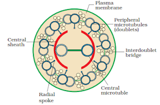
$\displaystyle \small \bullet$ Their core called the axoneme, possesses a number of microtubules running parallel to the long axis.
$\displaystyle \small \bullet$ The axoneme usually has nine pairs of doublets of radially arranged peripheral microtubules and a pair of centrally located microtubules.
$\displaystyle \small \bullet$ Such an arrangement of axonemal microtubules is referred to as the 9+2 array.
$\displaystyle \small \bullet$ The central tubules are connected by bridges and are also enclosed by a central sheath, which is connected to one of the tubules of each peripheral doublets by a radial spoke.
$\displaystyle \small \bullet$ Thus, there are nine radial spokes. The peripheral doublets are also interconnected by linkers.
$\displaystyle \small \bullet$ Both the cilium and flagellum emerge from centriole-like structure called the basal bodies.
$\displaystyle \small \bullet$ Cilia beats are asymmetrical with two distinct phases namely power effective phase and recovery phase.
$\displaystyle \small \bullet$ Flagella possess symmetrical beat with several undulations along its length.
CENTROSOME AND CENTRIOLES
$\displaystyle \small \bullet$ Centrosome is an organelle that generally have two cylindrical structures known as centrioles.
$\displaystyle \small \bullet$ Both the centrioles lie perpendicular to each other.
$\displaystyle \small \bullet$ They are made up of nine evenly spaced peripheral fibrils of tubulin.
$\displaystyle \small \bullet$ The central part of the centriole is also proteinaceous and called the hub, which is connected with tubules of the peripheral triplets by radial spokes made of protein.
$\displaystyle \small \bullet$ They are helpful in giving mechanical support, motility and maintenance of the shape of the cell.
$\displaystyle \small \bullet$ Help in the formation of cilia and flagella of the cells.


0 Comments